|
درباره دانش قلب
The heart is a muscular organ in most animals, which pumps blood through the blood vessels of the circulatory system. قلب یک ارگان عضلانی در بیشتر حیوانات است که خون را از طریق رگهای خونی سیستم گردش خون پمپ می کند. Blood provides the body with oxygen and nutrients, as well as assisting in the removal of metabolic wastes. خون اکسیژن و مواد مغذی بدن را فراهم می کند ، همچنین به دفع مواد زاید متابولیک کمک می کند. In humans, the heart is located between the lungs, in the middle compartment of the chest. در انسان قلب بین ریه ها ، در محفظه میانی سینه قرار دارد.
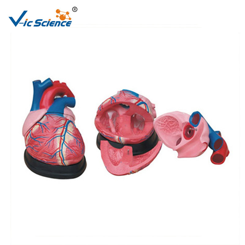
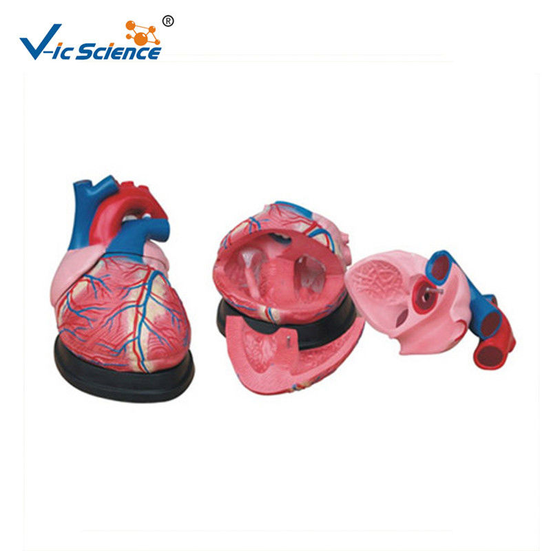
In humans, other mammals, and birds, the heart is divided into four chambers: upper left and right atria and lower left and right ventricles.Commonly the right atrium and ventricle are referred together as the right heart and their left counterparts as the left heart. در انسانها ، سایر پستانداران و پرندگان ، قلب به چهار اتاق تقسیم می شود: دهلیز فوقانی چپ و راست و بطن های پایین و راست. . Fish, in contrast, have two chambers, an atrium and a ventricle, while reptiles have three chambers. در مقابل ، ماهی ها دارای دو اتاق ، یک دهلیز و یک بطن هستند ، در حالی که خزندگان دارای سه اتاق هستند. In a healthy heart blood flows one way through the heart due to heart valves, which prevent backflow. در قلب سالم خون به دلیل دریچه های قلب یک طرف از طریق قلب جریان می یابد ، که مانع از بازگشت جریان خون می شود. The heart is enclosed in a protective sac, the pericardium, which also contains a small amount of fluid. قلب در یک کیسه محافظ ، پریکارد ، محصور می شود که حاوی مقداری مایعات نیز هست. The wall of the heart is made up of three layers: epicardium, myocardium, and endocardium. دیواره قلب از سه لایه تشکیل شده است: اپیکارد ، میوکارد و غدد درون ریز.
The heart pumps blood with a rhythm determined by a group of pacemaking cells in the sinoatrial node. قلب با ریتمی که توسط گروهی از سلولهای ضربان ساز در گره سینواتریال تعیین می شود ، خون را پمپ می کند. These generate a current that causes contraction of the heart, traveling through the atrioventricular node and along the conduction system of the heart. اینها جریان ایجاد می کنند که باعث انقباض قلب می شود ، از طریق گره دهلیزی و در امتداد سیستم هدایت قلب حرکت می کند. The heart receives blood low in oxygen from the systemic circulation, which enters the right atrium from the superior and inferior venae cavae and passes to the right ventricle. قلب خون کم خون از اکسیژن از گردش سیستمی دریافت می کند که از دهانه ورید برتر و تحتانی وارد دهلیز راست می شود و به بطن راست می رود. From here it is pumped into the pulmonary circulation, through the lungs where it receives oxygen and gives off carbon dioxide. از اینجا به گردش خون ریوی ، از طریق ریه هایی که اکسیژن دریافت می کند و دی اکسید کربن خاموش می کند ، پمپ می شود. Oxygenated blood then returns to the left atrium, passes through the left ventricle and is pumped out through the aorta to the systemic circulation−where the oxygen is used and metabolized to carbon dioxide. خون اکسیژن یافته به دهلیز چپ باز می گردد ، از بطن چپ عبور می کند و از طریق آئورت به گردش سیستمی می رسد - جایی که اکسیژن استفاده می شود و به دی اکسید کربن متابولیزه می شود. The heart beats at a resting rate close to 72 beats per minute. ضربان قلب با سرعت استراحت نزدیک به 72 ضربان در دقیقه. Exercise temporarily increases the rate, but lowers resting heart rate in the long term, and is good for heart health. ورزش به طور موقت سرعت را بالا می برد ، اما ضربان قلب استراحت را در طولانی مدت کاهش می دهد و برای سلامت قلب مفید است.

ساختار
The human heart is situated in the middle mediastinum, at the level of thoracic vertebrae T5-T8. قلب انسان در مدیاستن میانی ، در سطح مهره های قفسه سینه T5-T8 قرار دارد. A double-membraned sac called the pericardium surrounds the heart and attaches to the mediastinum. یک کیسه دو غشایی به نام پریکارد قلب را محاصره کرده و به مدیاستین وصل می کند. The back surface of the heart lies near the vertebral column, and the front surface sits behind the sternum and rib cartilages. سطح پشتی قلب در نزدیکی ستون مهره قرار دارد و سطح جلوی آن در پشت غضروف استرنوم و دنده قرار دارد. The upper part of the heart is the attachment point for several large blood vessels—the venae cavae, aorta and pulmonary trunk. قسمت فوقانی قلب محل اتصال چندین رگ خونی بزرگ است - ورید گاوی ، آئورت و تنه ریوی. The upper part of the heart is located at the level of the third costal cartilage. قسمت فوقانی قلب در سطح غضروف گرانشی سوم قرار دارد. The lower tip of the heart, the apex, lies to the left of the sternum (8 to 9 cm from the midsternal line) between the junction of the fourth and fifth ribs near their articulation with the costal cartilages. نوک تحتانی قلب ، اوج ، در سمت چپ ساق پا (8 تا 9 سانتی متر از خط میانی) بین محل اتصال دنده های چهارم و پنجم در نزدیکی مفصل آنها با غضروف های گرانشی قرار دارد.
The largest part of the heart is usually slightly offset to the left side of the chest (though occasionally it may be offset to the right) and is felt to be on the left because the left heart is stronger and larger, since it pumps to all body parts. بزرگترین قسمت قلب معمولاً کمی در سمت چپ قفسه سینه جبران می شود (هرچند گاهی ممکن است در سمت راست جبران شود) و احساس می شود که در سمت چپ قرار دارد زیرا قلب چپ قوی تر و بزرگتر است ، زیرا برای همه پمپ می شود. اعضای بدن. Because the heart is between the lungs, the left lung is smaller than the right lung and has a cardiac notch in its border to accommodate the heart. از آنجا که قلب بین ریه ها قرار دارد ، ریه چپ از ریه راست کوچکتر است و دارای یک شکاف قلبی در مرز خود برای جای دادن قلب است. The heart is cone-shaped, with its base positioned upwards and tapering down to the apex. قلب مخروطی شکل است و پایه آن به سمت بالا قرار گرفته و به سمت قله فرو رفته است. An adult heart has a mass of 250–350 grams (9–12 oz). قلب بزرگسال بالغ جرم 250-350 گرم (9 تا 12 اونس) دارد. The heart is often described as the size of a fist: 12 cm (5 in) in length, 8 cm (3.5 in) wide, and 6 cm (2.5 in) in thickness, although this description is disputed, as the heart is likely to be slightly larger. قلب اغلب به اندازه یک مشت توصیف می شود: طول 12 سانتی متر (5 اینچ) ، 8 سانتی متر (3.5 اینچ) عرض و ضخامت 6 سانتی متر (2.5 اینچ) ، اگرچه این توضیحات مورد اختلاف است ، زیرا قلب احتمال دارد کمی بزرگتر باشد Well-trained athletes can have much larger hearts due to the effects of exercise on the heart muscle, similar to the response of skeletal muscle. ورزشکاران خوب آموزش دیده می توانند به دلیل تأثیرات ورزش بر عضله قلب ، شبیه به پاسخ عضله اسکلتی ، قلب بسیار بیشتری داشته باشند.
|
 پیام شما باید بین 20 تا 3000 کاراکتر باشد!